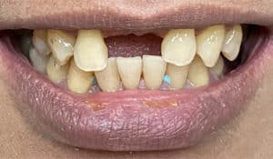
Case Details
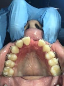
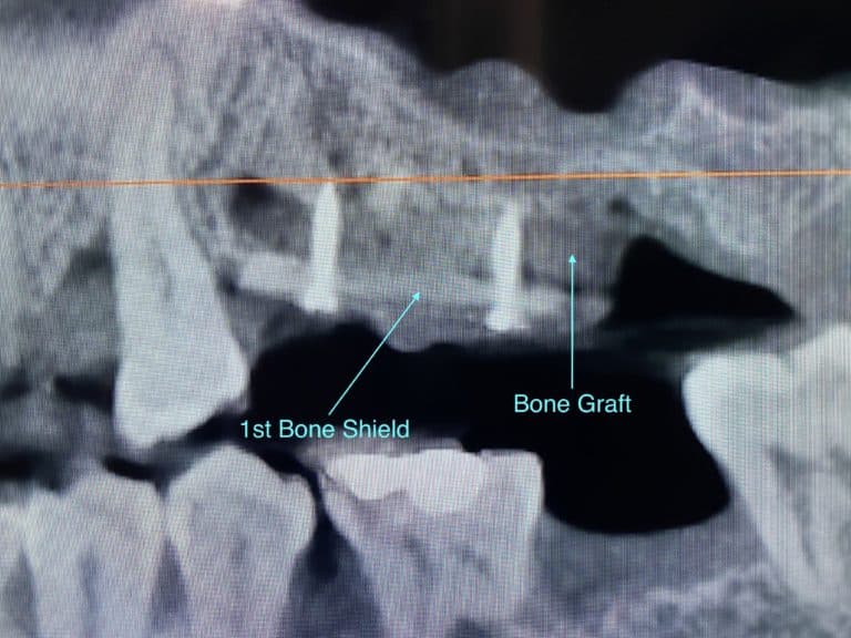
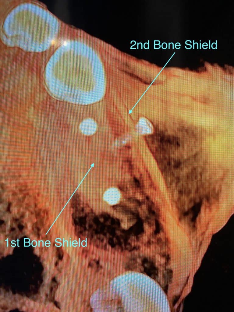
A patient lacks the necessary bone to support dental implants in the upper left quadrant and therefore requires a bone augmentation procedure. A bone bone block graft is harvested from the patient’s lower jaw (donor site) and 2 bone block veneers (aka bone shields, bone shells, bone blades or bone plates) are created from it. In addition, Bone chips and bone particulates are harvested from the patient as well and are all placed into the upper left quadrant (recipient site) along with growth factors. After extracting a hopeless tooth and another tooth with root tip fragments, a vertical augmentation of the ridge is implemented by placing and fixating the 1st bone shield at the desired height for the desired new bone growth. The 2nd bone shield is placed and fixated sideways or perpendicular to the 1st bone shield to provide a protective barrier for the autogenous bone graft during the healing phase. The area is then allowed to heal for a minimum of 4 months before the dental implants are placed.
Before
After

Before
After
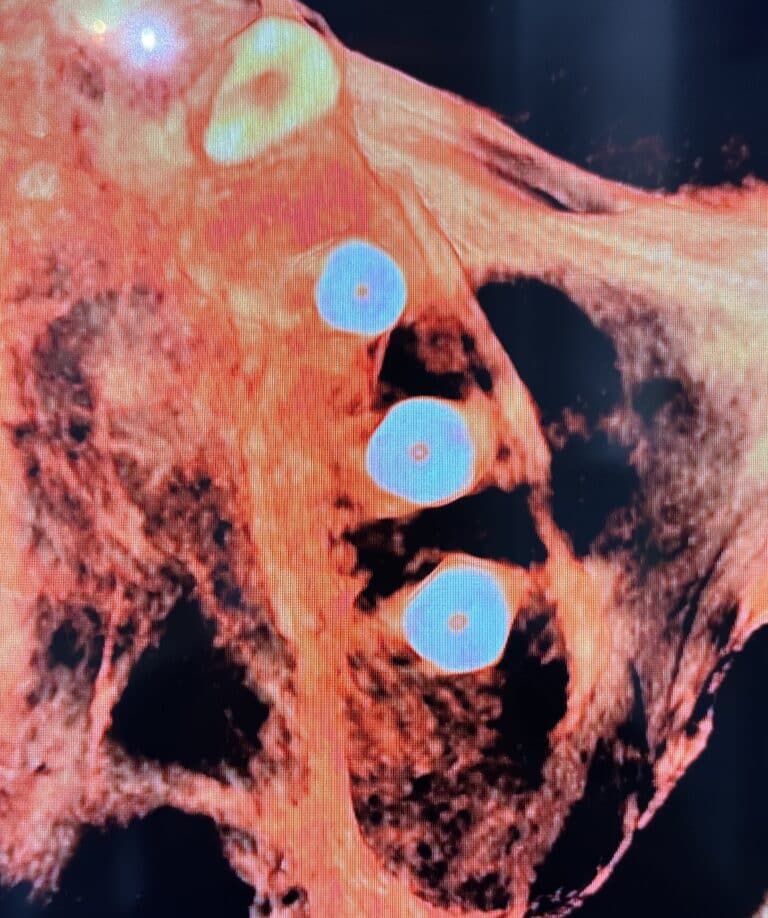
Description: CBCT inverted axial view (underbelly view) of the bone shields stabilized with fixation screws for the containment of the harvested bone graft.
Before
After
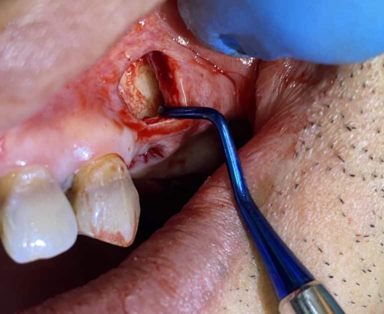
Description: A tunnel technique is utilized where there will be no flaps created. Only from this one incision is where the recipient site for the bone is prepared and all of the contents will be inserted. This method will increase the overall success of the split bone block (SBB) technique. After the completion of the procedure, the patient is allowed to heal for 4-6 months before the surgical site is ready for the placement of implants.
Before
After
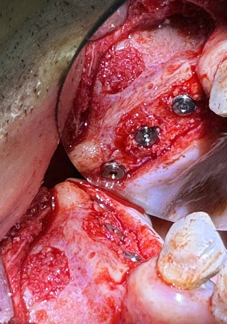
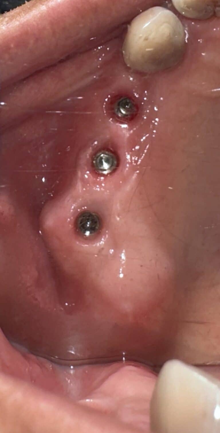
Description: After 4-6 months of healing has occurred, the surgical site is completely exposed and 3 implants are placed into the designated areas. Simultaneously, a sinus lift via the lateral window approach is performed along with BSM (bone substitute material) bone graft is added to further increase the height of the bone internally within the sinus cavity so that longer implant sizes could be placed.
Before
After
Description: CBCT inverted axial view of the 3 placed implants within newly formed bone.
Before
After
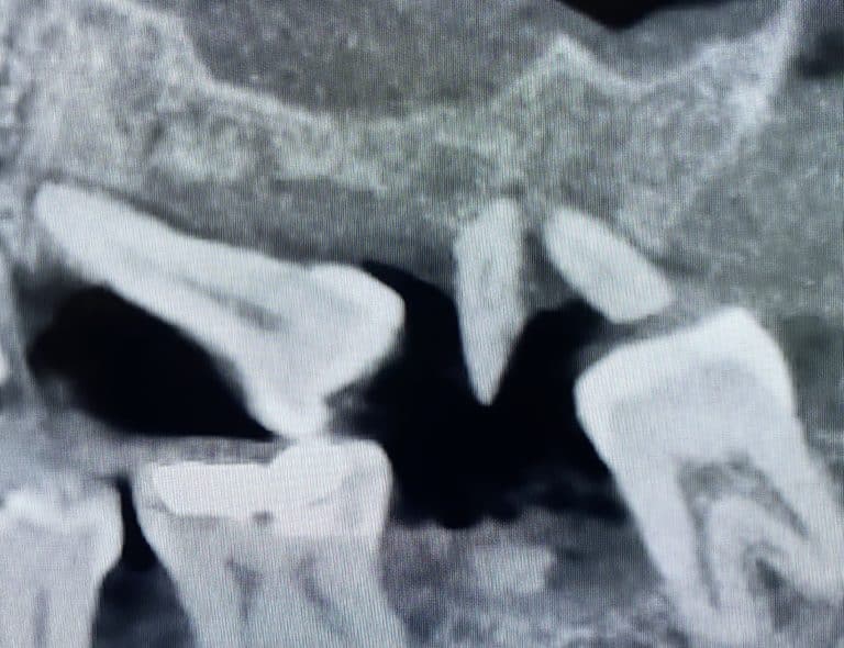
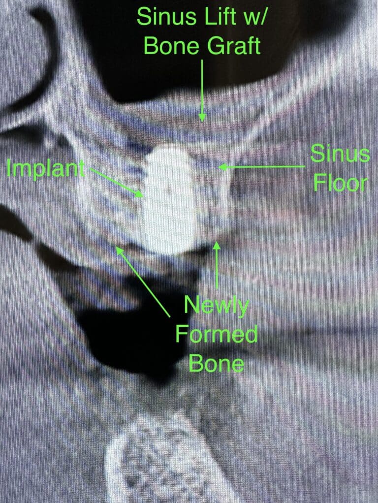
Description: A cross-sectional X-ray image from the most deficient area of 3 to 4 mm (initially) showing the harvested (transplanted) bone from the jaw has healed thus giving a new total alveolar ridge height of approximately 9 to 10 mm at this particular site. At the same time, an external sinus elevation was performed so that additional bone grafting material can be added on top of the sinus floor. As a result, a grand total of 15 to 16 mm of alveolar ridge height will be achieved thus allowing the option of placing longer implants if desired.
Before
After
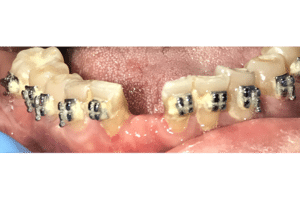
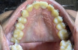
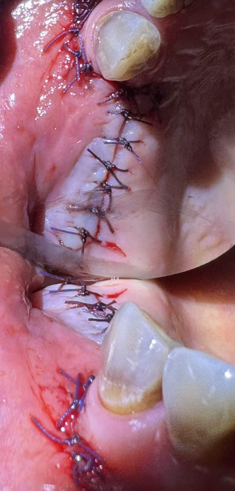
Description: The margins of the flap are reapproximated and sutured together with single interrupted sutures. The patient will return in 6 weeks to begin the process of restoring the area with prosthetics.
Before
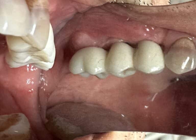
After
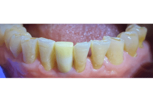
Description: After the designated time for healing has occurred, the implants are then ready to be connected to an oral prosthesis.
Before
After